Crohn’s disease (CD) is characterized by chronic, non-specific granulomatous inflammation that can involve any part of the gastrointestinal tract [1]. Around 22–44% of patients with CD will develop its cutaneous manifestation during lifetime [2]. Skin lesions are classified under three categories. The first group is related to the direct spread of the inflammatory process through the continuity from the gastrointestinal tract to the surrounding skin. The second group includes reactive dermatological conditions showing a strong correlation with CD (e.g. pyoderma gangrenosum, erythema nodosum). The third group is the extra-intestinal cutaneous form of the disease, referred to as ‘metastatic CD’ (MCD), in which skin lesions develop in locations not directly adjacent to the gastrointestinal tract but their histology shows non-caseating granulomatous infiltration typically observed in the intestinal variant of CD [3, 4]. In 70% of adults, MCD develops long after the diagnosis of CD, but it may pose a major diagnostic challenge in patients without gastrointestinal symptoms [5]. It should be also noted that the development of skin lesions, especially in case of MCD, does not correlate with disease activity in the gut [6].
We present the case of a female with MCD and highlight the dermoscopic and reflectance confocal microscopic (RCM) findings in this rare entity.
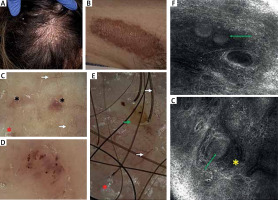
A 21-year-old woman with CD diagnosed in childhood was referred to the out-patient clinic for evaluation of a slowly growing, slightly pruritic erythematous plaque on the hairy scalp of 1-year duration (Figure 1 A). A similar erythematous-brownish infiltrated lesion developed in the right groin 5 years prior to referral (Figure 1 B). The patient had previously been consulted by two dermatologists who initially diagnosed psoriasis based on the clinical picture alone. The CD was in remission, the patient did not report any gastrointestinal symptoms and she was on maintenance therapy with sulfasalazine.
Figure 1
A – Clinical presentation – erythematous plaque on the hairy scalp with fine whitish scaling and reduced hair density; B – Clinical presentation – erythematous-brownish infiltrated plaque in the right groin; C – Non-polarized dermoscopy of the plaque in the groin showing linear branched vessels of irregular distribution (white arrows), fine white scales (red asterisk) and focally distributed pink-to-red structureless areas (black asterisk) over yellow-to-orange background (10×); D – Clustered irregularly coiled (linear curved) vessels under non-polarized dermoscopy (30×); E – Non-polarized dermoscopy of the plaque on the scalp showing multiple irregular linear branched vessels (white arrows), white scales (red asterisk) and perifollicular pustules (green arrow); F – Reflectance confocal microscopy of the lesion in the groin showing hyporeflective ovoid structures (green arrow) in the papillary dermis (VivaScope® 1500, MAVIG GmbH); G – Reflectance confocal microscopy of the plaque on the scalp showing a hyporeflective ovoid structure (green arrow) surrounded by linear vessels (yellow asterisk) (VivaScope® 1500, MAVIG GmbH)

On admission, non-polarized contact dermoscopy of both plaques showed presence of well-focused linear branched vessels and diffuse yellow-to-orange structureless areas (Figures 1 C–E). Several small erosions covered with crusts and fine white scales were present. In addition, pink-to-red structureless areas of focal distribution were noted within the lesion in the groin (Figure 1 C). Clustered irregularly coiled vessels and red haemorrhagic globules were also present (Figure 1 D). Within the plaque on the scalp, several perifollicular round yellowish structures, corresponding to pustules, were observed. Hair thinning and reduced number of hair follicles, without hair shaft abnormalities, was noted (Figure 1 E).
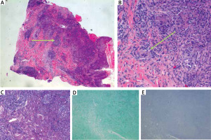
Visualization with RCM (VivaScope® 1500, MAVIG GmbH) showed hyporeflective ovoid structures in the papillary dermis, and increased vascularization (Figures 1 F, G). Both dermoscopic and RCM findings raised the suspicion of a granulomatous disorder, while RCM was also helpful in choosing the biopsy site. Histopathological examination of both plaques showed presence of a profuse inflammatory infiltrate in the dermis, composed mainly of lymphocytes, histiocytes, and a few non-caseating granulomas with multinucleated giant cells (Figure 2 A). Ancillary stainings, including Periodic Acid Schiff (PAS), Grocott-Gomori silver stain for fungal organisms and Treponema, and ABS staining for Mycobacterium, were all negative (Figures 2 B–D). Based on the clinical details, histopathological examination and additional stainings, the diagnosis of MCD was made.
Figure 2
A – Histopathology showing presence of granulomatous infiltrate with numerous lymphocytes (green arrow) and a few non-caseating granulomas with multinucleated giant cells in the dermis (40×; hematoxylin and eosin staining); B – Non-caseating granuloma with a multinucleated cell (green arrow) (200×; hematoxylin and eosin); C – negative Periodic Acid Schiff (PAS) for fungi; D – negative Grocott-Gomori silver stain for fungi and Treponema; E – negative AFB staining for Mycobacterium

Dermoscopy has already been demonstrated to be a useful tool for the preliminary diagnosis of several cutaneous granulomatous conditions, including sarcoidosis, leishmaniasis, granuloma annulare, necrobiosis lipoidica and granulomatous rosacea [7]. Although granulomatous diseases show some similarities in dermoscopic examination, such as presence of orange or yellow-to-orange structureless areas and arborizing vessels, there are some clues that may facilitate differential diagnosis. In necrobiosis lipoidica, the structureless areas display a yellower hue, probably due to the presence of lipid deposits in the dermis [8]. In granuloma annulare, the orange-to-yellow structureless areas may not be visible, especially in the interstitial variant of the disease or when granulomas are located in deep dermis. In granulomatous rosacea, the structureless areas show predominantly focal distribution and are accompanied by linear vessels arranged in a polygonal pattern [8]. On the other hand, follicular plugs, commonly displaying a tear-drop shape, and white starburst-like pattern are highly characteristic of cutaneous leishmaniasis [8]. In the presented patient, we observed distinct dermoscopic features, namely pink-to-red structureless areas of focal distribution and clustered coiled vessels. In addition, subclinical pustules (visible under dermoscopy but not with naked eye) were present within the plaque on the scalp. These findings may constitute specific dermoscopic findings for MCD. However, studies on larger groups of patients are needed.
Although MCD can affect any area, it has been predominantly observed on the extremities or in the genital region [4, 5]. The involvement of the scalp is extremely rare; therefore, we would like to highlight the trichoscopic findings in particular. Data on the potential application of RCM in cutaneous granulomatous disorders are predominantly based on single case reports [9–11]. To the best of our knowledge, dermoscopic or RCM features of MCD have not been reported in the literature yet. In the presented patient, dermoscopic findings were suggestive of the granulomatous condition. In addition, RCM enabled visualization of sparse granulomas. On histopathology, non-caseating granulomas are less well-formed and harder to find in MCD when compared to e.g. sarcoidosis. For that reason, RCM-guided biopsy may be of help.


