


Linear immunoglobulin A (IgA) disease (LAD), also known as linear IgA bullous dermatosis (LABD), is an autoimmune mucocutaneous disease with an estimated annual incidence of 0.2–2.3 cases per million/population. It may occur in adults and children, and is known as chronic bullous disease of childhood (CBDC) [1].
LABD is an idiopathic pathology caused by increased circulating IgA anti-basement membrane zone antibodies against the lamina lucida [2]. Presence of linear IgA deposits at the dermoepidermal junction observed by applying direct immunofluorescence is mandatory to confirm the diagnosis (gold standard) [1, 3].
Association with autoimmune pathologies or certain drugs have been described (representing 37.5% of LABD in adults, with an atypical, extensive and more severe behaviour than the idiopathic form) [3]. Clinical manifestation includes polycyclic groupings of bullae with central crusting called strings of pearls as the most characteristic clinical finding, especially in children and less frequently in adults. Cases mimicking Steven-Johnson syndrome (SJS) or toxic epidermal necrolysis (TEN) have been described [4].
We present the case of an 89-year-old woman who developed LABD during piperacillin-tazobactam therapy. The patient developed intertriginous lesions after receiving antibiotic therapy just as described in the previous literature [5]. Medical personal history highlights a mild cognitive impairment, type 2 diabetes mellitus, chronic kidney disease, atrial fibrillation and treatment with furosemide, metformin, edoxaban and felodipine/metoprolol.
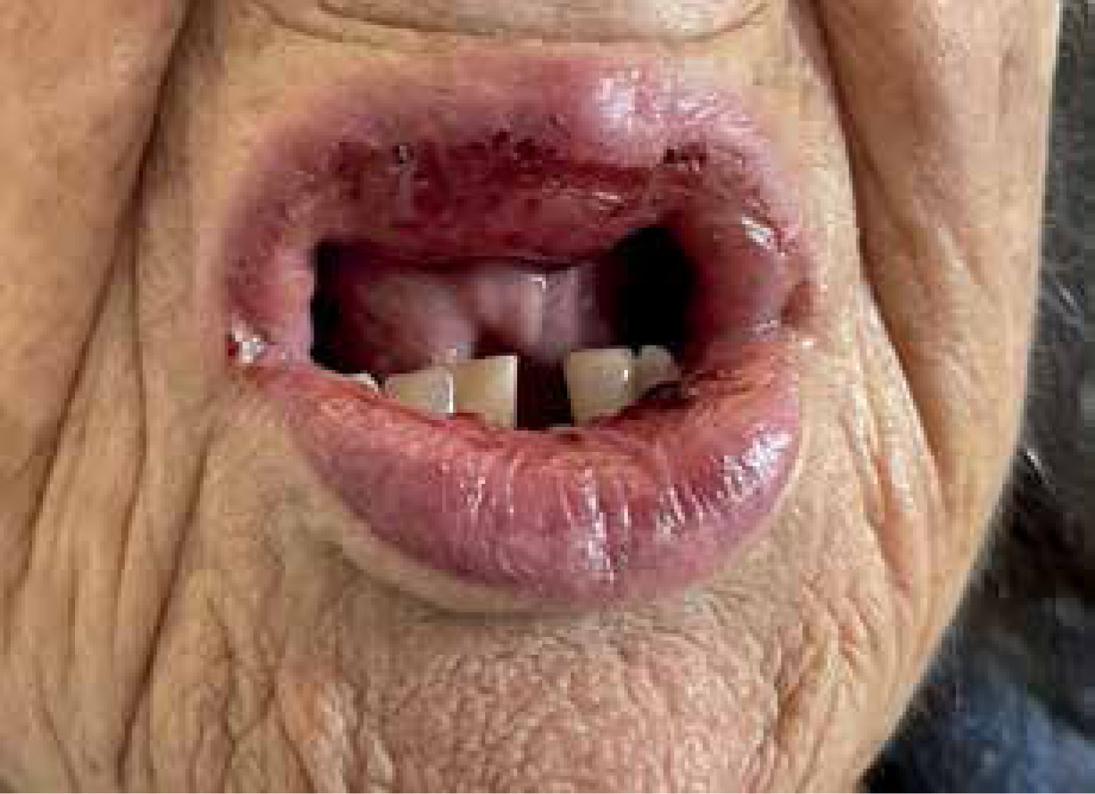
Admission criteria was secondary to acute cholecystitis and bacteriemia due to Escherichia coli, so piperacillin-tazobactam therapy was initiated. On the eleventh day of treatment, erythematous papular lesions and vesicles with no desquamation began to appear on the submammary fold. She also presented with vesicles and bullae on an erythematous base on both palms, erythema on soles and papular and bullous lesions on the inguinal and proximal region of lower limbs. Additionally, she presented with generalized itching, angioedema and bullous lesions on lips, lingual and soft palate mucosa which became painful by their rupture (Figure 1).
The allergology department was notified and evaluated the patient with the diagnosis of possible SJS or autoimmune blistering disease secondary to piperacillin-tazobactam. The antibiotic was discontinued and no other antimicrobials were required due to resolution of the infection. Treatment with intravenous corticosteroids and antihistamines was started, as well as zinc-sulfate was applied to the blistered areas, resolving symptoms in 6 days.
Blood and biochemistry analysis was normal (eosinophils 2.6%, no liver or renal involvement), Cytomegalovirus IgG (169), Epstein-Barr virus IgG (> 750), EBV-nuclear antigen (> 600); and Herpes-simplex virus type 1 and 2 IgG were positive with negative IgM. Parvovirus-B19 and human herpesvirus 6 were negative.
Skin lesion biopsy on the thigh showed histopathological findings suggestive of LABD in adults: thinned epidermis with spongiosis and isolated necrotic keratinocytes was observed under which there was a subepidermal blister with neutrophils and eosinophils within it and in the concomitant papillary dermis. Direct immunofluorescence showed intense linear deposits of IgA along the basement membrane, being negative for IgG, IgM, C3, fibrin and C1Q.
Although hematoxylin-eosin findings may be similar in SJS, dermatitis herpetiformis and LAD, linear-IgA deposit is characteristic of the last 2 entities, ruling out SJS in this case. The pathologist reported that differential diagnosis between the last 2 entities was almost impossible, but clinical findings and the extensive existing bibliography that documents association of LAD in adults with certain drugs, suggest this possibility as the most appropriate diagnosis in this context and her positive development after discontinuing the drug.
In order to provide the patient with safe alternatives within β-lactam antibiotics, intraepidermal and intradermal skin tests were performed with cefuroxime, ceftriaxone and ceftazidime, obtaining negative results in immediate and delayed reading. In a subsequent hospital admission, the patient received ceftazidime without any incident. Studying aminopenicillins was avoided given the severity of the pathology, the age and baseline situation of the patient.
LABD is a rare entity. There is a higher incidence in childhood (6 months–10 years) and in the sixth decade of life [6]. Our patient is an elderly woman in her late eighties and few cases have been described in such elderly patients.
Risk factors for developing LABD include associations with the human leukocyte antigen (HLA) B8, HLA-DR3, HLA-Cw7, HLA-DQ2 and tumor necrosis factor-2 allele, but more studies are needed to determine the involvement of drugs and these genetic risk factors [7].
Classical presentation in children consists of plaques and polycyclic or annular papules with blisters on the edges (“string of pearls”) that appear first around the eyes and mouth, then on the lower abdomen, genitals, thighs, buttocks, wrists, and ankles. In adults, the “string of pearls” is less frequent and appears on the trunk, extremities and head. Mucosal involvement appears in both cases but in adults it is present in 80%. Itching may be non-existent, mild or severe.
If the disease affects the adult population, the drug-induced form should be reviewed [8–10].
Vancomycin is the most frequently implicated drug, being involved in 50% of cases. Other drugs including β-lactams, sulfonamides, angiotensin-converting enzyme inhibitors (ACEI), non-steroidal anti-inflammatory drugs (NSAIDs), phenytoin, furosemide, allopurinol, amiodarone and atorvastatin have also been described [1].
Rapid diagnosis is essential, as well as establishing an adequate causal relationship with the drug involved. Symptoms usually begin within the first month of treatment. Discontinuation of the culprit drug is mandatory. Dapsone, sulfonamides, colchicine, methotrexate, cyclosporine, topical and oral corticosteroids, or intravenous immunoglobulins (IVIG) have been described as therapy [1].
Our patient underwent piperacillin-tazobactam treatment for 11 days until symptoms appeared. In another case due to piperacillin-tazobactam described in the literature, symptoms began 3 days following the start of antibiotic treatment. Penicillins are the least frequent involved antibiotics [5]. In both cases, intertriginous areas were affected. Resolution in 1 week was achieved after discontinuing piperacillin-tazobactam and starting treatment.
The most important thing in this pathology is obtaining a biopsy of the injured skin area (obtained from a perilesional skin area [4]) for staining with hematoxylin-eosin and immunofluorescence.
Early recognition and review of the drugs taken by the patient is important in order to obtain a sooner recovery since the pharmacological origin in adults is the most frequent.
To conclude, given the low frequency of this pathology in the general population, a suspected diagnosis is necessary, and for that, this clinical entity must be known.