


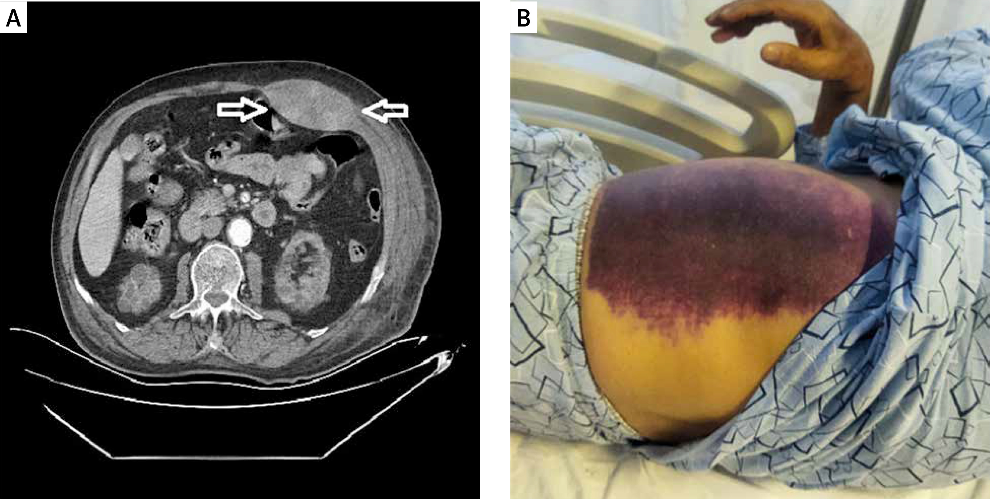
A 74-year-old male patient was admitted to our emergency department with sudden onset of chest pain. His medical history included hypertension and diabetes mellitus. On arrival, cardiac enzyme levels were increased, and electrocardiography revealed ischemic findings. A significant lesion in the left anterior descending artery was observed during the emergency coronary angiography. The percutaneous coronary intervention was successfully performed using intravenous unfractionated heparin 100 μg/kg and oral ticagrelor 180 mg loading dose. On the second day after the procedure, the patient’s urine output stopped, blood creatinine levels increased, and hemodialysis was performed for acute renal failure. During hemodialysis via the right subclavian vein, the patient developed a sudden onset of left abdominal pain. Abdominal distension and tenderness were observed, and Carnett’s sign was positive. Abdominal computed tomography (CT) revealed rectus sheath hematoma (Figure 1 A). In the following days, diffuse ecchymosis was observed in the left abdominal region (Figure 1 B). On the 7th day, the patient’s pain and hematoma regressed, the urine output was restored, and successful discharge was achieved.
Figure 1
A – Abdominal CT image of rectus sheath hematoma (with arrow). B – Diffuse ecchymosis of the skin in the left abdominal region
Rectus sheath hematoma is an uncommon cause of acute abdominal pain that typically originates from tears in the rectus abdominis muscle or ruptures of the abdominopelvic arteries and their branches, particularly when anticoagulant medication is used [1]. Coughing, straining, aging, abdominal surgery, obesity, subcutaneous injections, anticoagulant therapy, and trauma are the most frequent risk factors [2]. While the majority of cases respond well to conservative treatment, an interventional strategy may be required in select instances.